Fiction
Pharmaceutical Microbiology
by Amal Bobby
3 chapters16 min read
Chapter 1
Foundations of Microbiology
The lecture hall hummed with the low murmur of students settling into their seats, the scent of chalk dust and stale air lingering near the back rows. Arjun shifted in his chair, notebook open but untouched, feeling the weight of unfamiliar terms press against his temples like a tightening band. Pharmaceutical Microbiology. The words on the syllabus had looked innocuous enough, but now, as Professor Meera stepped toward the podium, they seemed to carry a gravity he wasn’t prepared for.
"Good morning," she began, her voice calm and clear, cutting through the chatter. "Before we dive into stains and sterilizers, let’s ask: why does microbiology matter to a pharmacist?"
A few hands lifted hesitantly. Arjun kept his eyes down, tracing the edge of his page. He knew microbes were tiny living things—bacteria, viruses, fungi—but what did that have to do with dispensing tablets or checking prescriptions?
Professor Meera smiled, not unkindly. "Think of a drug not just as a chemical compound, but as a product that travels from factory to patient. At every step, it’s vulnerable. Not to breaking, but to being invaded."
She paused, letting the idea settle. "Microbes don’t need much. A drop of moisture, a speck of dust, a moment of exposure—and they can multiply. In a syrup, they ferment sugars and produce gas. In an ointment, they break down emulsions. In an injectable, they can endanger a life."
Arjun’s breath hitched. He pictured the IV bag his aunt had received during her hospital stay—clear, hanging silently by her bedside. What if it hadn’t been sterile? What if, instead of healing, it had introduced something harmful?

Professor Meera paced slowly, her gestures deliberate. "Pharmaceutical microbiology is the science that protects the integrity of medicines. It ensures that what we manufacture is not only potent but pure. That a tablet doesn’t carry pathogens that could cause infection. That a cream doesn’t grow mold that could irritate skin. That a vial of insulin is safe to inject into a child’s bloodstream."
She stopped and faced the class. "Contamination isn’t always visible. You won’t see a cloud of bacteria in a liquid antibiotic. But if it’s there, and it’s pathogenic, the consequences can be severe—sepsis, organ failure, even death. Our role isn’t just to formulate drugs; it’s to ensure they don’t harm the very people they’re meant to heal."
Arjun’s fingers tightened around his pen. The abstract fear he’d felt moments ago sharpened into something concrete. This wasn’t just about memorizing names or drawing diagrams. It was about responsibility. About the quiet, unseen work that kept patients safe.
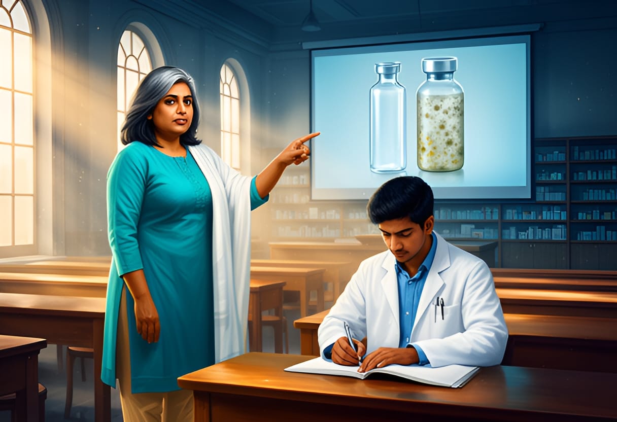
Professor Meera clicked the remote, and a slide appeared: two vials side by side. One labeled sterile, the other showing cloudy growth after incubation. "This," she said, "is why we test. Why we control environments. Why we validate processes."
Arjun exhaled, slowly. The fog in his mind began to lift—not all at once, but enough to see a path forward. He picked up his pen and wrote, not a definition, but a question: How do we stop them?
He would learn the answer soon enough. For now, the weight of the knowledge felt less like a burden and more like a purpose.
Professor Meera advanced to the next slide, which displayed a branching diagram labeled with terms Arjun had only glimpsed in his textbook—Gram-positive, Gram-negative, cocci, bacilli, spore-formers. "Classification," she said, "isn’t just about naming. It’s about predicting behavior. In pharmacy, we focus on bacteria because they’re the most common contaminants in non-sterile products and the hardest to control in sterile ones."
She tapped the screen. "We start with shape: cocci are spherical, bacilli are rod-shaped. Then arrangement—streptococci in chains, staphylococci in clusters. After that, Gram reaction, which tells us about cell wall structure and, importantly, how they respond to antibiotics and disinfectants. Spore formation is another key trait—some bacteria, like Bacillus and Clostridium, form highly resistant spores that can survive boiling and standard sterilization."
Arjun leaned forward, pen moving again. He sketched a tiny oval with a thick wall inside it—his version of an endospore. "So if a product is contaminated with spores, normal heating won’t kill them?"
"Exactly," Professor Meera said. "That’s why we use autoclaving—moist heat under pressure—for sterilization. Dry heat or filtration won’t touch spores reliably. And in syrups or creams, where we can’t sterilize the final product, we rely on preventing growth instead."
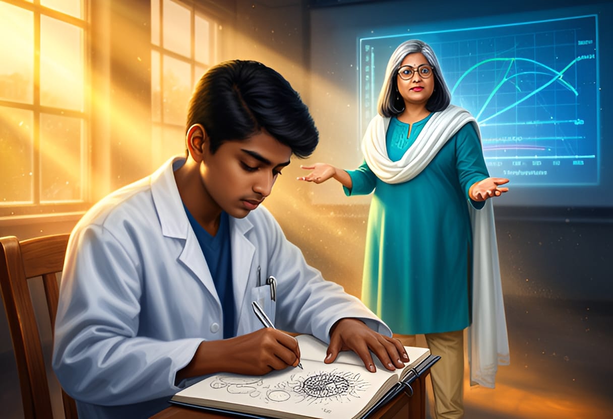
She shifted to a new slide showing a graph with temperature on the x-axis and growth rate on the y-axis. "Now, growth. Microbes don’t just appear—they need conditions. Think of it like planting a seed. You need the right soil, water, temperature. For bacteria, it’s nutrients, moisture, pH, temperature, and oxygen."
Arjun frowned. "But medicines aren’t soil. How do they get what they need?"
"Ah," Professor Meera said, smiling. "That’s the insight. Many pharmaceutical products—especially liquids, semisolids, and even some solids—contain water, sugars, proteins, or lipids that microbes can use. A cough syrup isn’t just medicine; it’s a nutrient broth if not preserved properly. The water activity, the pH, the presence of preservatives—all of these determine whether a contaminant can multiply."
She pointed to the graph. "Most pathogens thrive between 20 and 45 degrees Celsius—room to body temperature. That’s why accelerated stability testing often includes incubation at 37°C or higher. If a product shows no growth there, it’s less likely to spoil in tropical climates or during storage."
Arjun recalled his aunt’s insulin vial, stored in the refrigerator. "So cold slows them down?"
"Refrigeration inhibits growth but doesn’t kill," Professor Meera said. "Freezing may halt it, but thawing can revive survivors. That’s why we don’t rely on temperature alone. We combine strategies—low water activity, acidic pH, antimicrobial preservatives, protective packaging—to create multiple barriers."
A flicker of understanding passed through him. It wasn’t about eliminating microbes entirely in every case—it was about making the product inhospitable. Like building a dam not to stop rain, but to control where the water goes.
Professor Meera clicked again. The slide now showed three vials: one clear, one slightly hazy, one with visible mold. "Finally, not all microbes are equal in risk. Commensals—like some strains of Escherichia coli that live in our gut—may not cause disease, but their presence in a non-sterile product indicates a breach in hygiene. Pathogens, like Salmonella or Pseudomonas aeruginosa, are dangerous even in low numbers. And fungi? They’re tougher to kill, love acidic environments, and can ruin emulsions or produce toxins."
Arjun’s earlier question echoed in his mind: How do we stop them? Now he saw it wasn’t a single answer, but a layered strategy—knowing the enemy, understanding its needs, and cutting off its supply lines.
Professor Meera turned off the projector. "Remember this: in pharmaceutical microbiology, we’re not just fighting microbes. We’re designing products that deny them a chance to thrive. That’s how we keep medicines safe."
Arjun closed his notebook. The weight hadn’t vanished—but it had shifted. No longer a vague dread, it was now a challenge he could meet, one factor at a time. He looked up, ready for the lab. * The microbiology lab smelled of alcohol and agar. Rows of workstations held petri dishes, inoculating loops, and bottles of media glowing under ultraviolet lights. Sneha was already at their bench, labeling tubes with neat, precise script.
"You’re late," she said, not looking up. "Professor Meera’s waiting."
Arjun slipped on his lab coat, the familiar weight grounding him. "Sorry. Just… thinking."
"About the lecture?" Sneha asked, finally meeting his eyes. She tilted her head. "You look like you finally get it."
He smiled, small but real. "I think I do. It’s not about memorizing every bug. It’s about asking: what does it need to live? And then… not giving it to them."
Sneha nodded. "Exactly. Now, let’s see what grows when we do."

Chapter 2
Understanding Bacteria
Arjun adjusted the focus knob, the coarse then fine, until the blurred smudge resolved into distinct purple spheres clustered like grapes. "Staphylococcus," he murmured, more to himself than to Sneha. The coccus arrangement was unmistakable—irregular clusters, not chains or pairs.
"Good," Sneha said, not looking up from her own slide. "Now find the diplococci. They’ll be in pairs, side by side."
He scanned the field, pausing where two oval dots sat fused at their edges. "Neisseria-like," he said. "But these are Gram-positive, right? Thick peptidoglycan holds the crystal violet."
"Exactly," came Professor Meera’s voice from behind. Arjun startled, then steadied himself. She leaned in, peering over his shoulder at the eyepiece. "What you’re seeing isn’t just shape—it’s architecture. The cell wall determines how the stain behaves, and ultimately, how vulnerable the bacterium is to our drugs."

Arjun blinked. "So the purple color means it’s Gram-positive because the thick wall traps the dye?"
"Precisely." She straightened, gesturing to the bench where a laminated chart hung. "Gram-positive bacteria have a thick peptidoglycan layer—sometimes up to 90% of the cell wall. Gram-negatives? Thin peptidoglycan, but an outer membrane rich in lipopolysaccharides. That outer layer acts like a shield. It’s why many antibiotics that target cell wall synthesis—penicillins, vancomycin—work better on Gram-positives. The Gram-negatives keep them out."
Sneha tapped her slide. "Like Pseudomonas aeruginosa. We saw it in the injectables lecture. Highly resistant, partly because of that outer membrane."
Professor Meera nodded. "And that’s why we test susceptibility. Structure predicts response. A bacterium isn’t just a blob—it’s a fortress with specific weak points. Your job isn’t to memorize every fort, but to understand how they’re built so you know where to aim."
Arjun returned his eye to the microscope. The bacilli on the next slide were long and rod-shaped, some forming end-to-end chains. Bacillus, he thought. Or maybe Clostridium. He adjusted the diaphragm, increasing contrast. The spores—if present—would be invisible without special staining, but the vegetative cells were clear: straight rods with squared ends, arranged in singles or short chains.
"What about size?" he asked, lowering the scope. "Does that matter for filtration or drug penetration?"
"Absolutely," Professor Meera said. "Most bacteria range from 0.5 to 5 micrometers. Cocci tend to be smaller—0.5 to 1.5 µm. Bacilli can be longer, but width stays similar. That’s why 0.22-micron filters work for sterilizing solutions—they exclude virtually all bacteria, though not viruses or mycoplasma. Size, shape, wall composition—all are clues. In the lab, we use them to identify. In formulation, we use them to exclude."
Arjun turned the stage, bringing a cluster of streptococci into view—beaded chains of cocci. He recalled the earlier lecture: not all microbes are equal in risk. Some strains of Escherichia coli were harmless in the gut but dangerous in a non-sterile cream. Now, seeing the chains, he thought not just of identification, but of implication. If this were a contaminant in a syrup, its chain formation might affect how it settled or clung to particles. If it were Gram-negative, its outer membrane might resist certain preservatives.
He looked up. Sneha was watching him, waiting.
"I’m seeing it now," he said. "It’s not just what they are. It’s how they’re made—and what that means for whether they live or die in our products."
Professor Meera smiled faintly. "Then let’s see what grows when we give them a chance." * Later, after they had heat-fixed slides and applied the primary stain, Arjun watched as the crystal violet flooded the slide. The bacteria absorbed the dye, turning deep purple. Then came the iodine—mordant, locking the color in. He held his breath during the decolorization step, alcohol trickling down the slide, terrified of over-washing and losing the signal.
"Too long and you’ll strip the Gram-positives bare," Sneha warned, her voice calm. "Too short and the Gram-negatives stay falsely purple."
He timed it, counting silently. When the runoff appeared clear, he stopped. The safranin counterstain followed—a faint pink wash. He wiped the slide gently, blew it dry, and placed it on the stage.
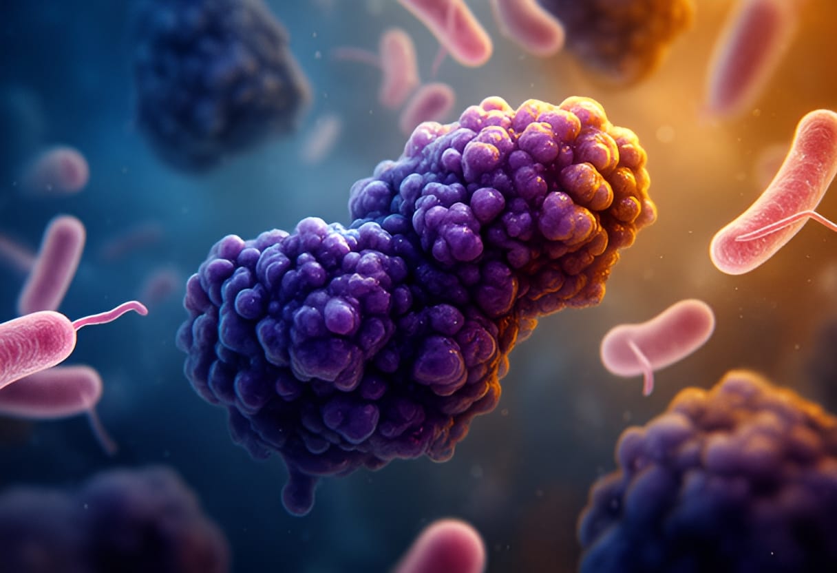
Under oil immersion, the Staphylococcus clusters shone purple, thick-walled and defiant. The Escherichia coli rods nearby were pale pink, their thin peptidoglycan unable to hold the primary stain against the alcohol’s assault.
Arjun exhaled. "So the wall isn’t just structure—it’s a gatekeeper. It decides what gets in… and what stays out."
Professor Meera, reviewing his slide, said, "And in pharmaceutical microbiology, we learn to build better gates."
The classroom lights hummed overhead as Professor Meera clicked to the next slide—a grayscale electron micrograph of a bacterium split open to reveal a dense, oval structure nestled within its cytoplasm. "This," she said, tapping the image, "is an endospore. Not all bacteria make them, but the ones that do—like Bacillus and Clostridium—can survive conditions that would kill ordinary vegetative cells: dry heat, radiation, desiccation, even many chemical disinfectants."
Arjun leaned forward. In the lab, he’d seen bacilli forming chains; now he understood why some of them worried formulators more than others. "So if a spore gets into a powder for reconstitution, it could lie dormant until the solution is made—and then germinate?"
"Exactly," Professor Meera said. "And once it germinates, you have a metabolically active bacterium capable of growth, toxin production, or both. That’s why sterility testing isn’t just about detecting what’s alive now—it’s about ensuring nothing can become alive later."
She advanced to a graph showing a sigmoidal curve labeled with lag, log, stationary, and death phases. "Bacterial growth isn’t instantaneous. In fresh medium, there’s a lag phase where cells adapt—synthesizing enzymes, repairing damage. Then comes exponential log phase, where biomass doubles predictably. Nutrients deplete, waste builds, and growth slows into stationary phase. Finally, if conditions don’t improve, death begins."
Sneha raised her hand. "But in a sealed vial of antibiotic, there shouldn’t be any nutrients, right? So how do they grow at all?"
"Good question," Professor Meera said. "Ideally, there are none. But real-world manufacturing isn’t perfect. Microscopic residues—water, sugars, proteins—can remain in equipment. A single spore in a moist niche can germinate, find enough to replicate, and before you know it, you have visible turbidity or gas production in a solution meant to be clear."
Arjun thought of the syrup fermentation Professor Meera had mentioned in lecture—the way unseen microbes could turn a sweet vehicle into a bubbling hazard. Now he saw it wasn’t just about contamination; it was about timing. A batch might pass initial bioburden testing if spores were still dormant, only to fail weeks later on the shelf. "So stability studies have to account for germination time?" he asked.
"They do," Professor Meera said. "Challenge testing introduces known spores or vegetative cells to simulate worst-case scenarios. We watch for growth over weeks, not just days. And in aseptic filling, we design processes to eliminate not just cells, but spores—hence the use of moist heat sterilization at 121°C for 15 minutes, which achieves a defined lethality against even the most resistant spores like those of Clostridium botulinum."
The next slide showed a biofilm: a forest of bacterial cells encased in a glistening matrix, adhered to a plastic surface. "This," Professor Meera said, "is how bacteria survive in water systems, on tubing, in valve seats. The extracellular polymeric substance shields them from biocides and shear forces. A biofilm can shed planktonic cells continuously—like a slow leak of contamination."
Sneha frowned. "So even if you flush the system, the biofilm stays?"
"Unless you remove it mechanically or treat it with agents that penetrate the matrix," Professor Meera said. "That’s why sanitization cycles in pharmaceutical water systems include oxidants like peroxide or peracetic acid, and why periodic hot water sanitization at 80°C is used—not to sterilize, but to disrupt biofilm integrity."
Arjun’s mind connected the dots. In sterile manufacturing, it wasn’t enough to kill free-floating cells. You had to address the reservoirs where bacteria hid and persisted. "So when we talk about aseptic technique, we’re not just preventing introduction—we’re also preventing growth from what’s already there?"
"Precisely," Professor Meera said. "Environmental monitoring isn’t just about air and surfaces. It’s about trending. Finding a single Bacillus spp. in a grade C area might be an anomaly. Finding it repeatedly? That suggests a niche—a crack in a gasket, a spill under a bench—where spores are protected and periodically dispersed. Your role as a pharmacist isn’t just to react to contamination, but to anticipate where it could take hold."
She turned off the projector. The room dimmed to the glow of the emergency exit sign. "Remember: a bacterium’s physiology dictates its danger. Its growth phase tells you when it’s most vulnerable. Its survival structures tell you how hard it is to eradicate. And its ability to form biofilms or spores tells you where to look—not just in the product, but in the process."
Arjun gathered his notes, the weight of the lecture settling not as anxiety, but as purpose. He hadn’t just learned how bacteria lived—he’d learned how they endured. And in that endurance lay the challenge: to design systems so rigorous, so attentive to the hidden life of microbes, that no spore, no biofilm cell, could slip through.
As he packed his bag, Sneha nudged him. "You okay?"
He smiled, real this time. "Yeah. I think I’m starting to see the whole picture—not just the bug, but the battlefield." * Later, in the quiet of the library, Arjun sat alone with a textbook open to a chapter on bacterial toxins. He’d spent the morning reviewing growth curves and sporulation conditions, but now he turned to pathogenicity—not just how bacteria survived, but how they harmed.
The text described exotoxins: proteins secreted by live cells that could disrupt cellular function at nanogram concentrations. Endotoxins, shed from the outer membrane of Gram-negatives upon lysis, could trigger fever, shock, even death if they entered the bloodstream. He thought of the injectables lecture—the vial that looked clear but carried pyrogens that could kill.
He remembered Professor Meera’s words about building better gates. Now he understood: it wasn’t enough to keep bacteria out. You had to ensure that even if fragments or products of their metabolism got in, they couldn’t cause harm. The gate had to screen not just for whole cells, but for the invisible weapons they carried.
He closed the book slowly. The responsibility wasn’t abstract anymore. It was in the filtration of a solution, the validation of a sterilization cycle, the testing for endotoxins in every batch meant for intravenous use. Each step was a decision point—a chance to stop a threat not just of contamination, but of injury.

Chapter 3
Staining Techniques for Identification
Arjun adjusted the focus knob on the microscope, squinting at the faint smudge on the slide. Beside him, Sneha tapped her pencil against her notebook, waiting. The microbiology lab smelled of alcohol and agar, the fluorescent lights humming overhead. He had just finished preparing a smear of Staphylococcus aureus and Escherichia coli, two standard strains provided for the Gram staining exercise. His hands still felt clumsy with the inoculating loop, the flame of the Bunsen burner a nervous presence in his peripheral vision.
"Remember," Professor Meera had said earlier, her voice cutting through the chatter of the lab, "the decolorization step is where most students lose the stain. Too little, and everything looks purple. Too much, and even the Gram-positives wash out. It’s a rinse, not a scrub."
Arjun had nodded, but now, holding the bottle of 95% ethanol, he hesitated. He tilted it over the slide, letting the alcohol flow. It rushed across the surface, and he pulled it away too quickly—just as he had the first time. The crystal violet drained away like water from a cracked cup. He stared. The slide was nearly blank.
"Again?" Sneha asked softly.
He swallowed. "I think I rushed it."
"Try again. Count to five slowly. Let it flow, don’t pour."
He re-stained the slide with crystal violet, waited a minute, rinsed gently with water, then flooded it with iodine. After another minute, he rinsed again. This time, when he brought the ethanol to the slide, he let it trickle down the edge, watching for the moment the purple began to fade from the edges. He counted: one Mississippi, two Mississippi… at five, he tipped the slide upright and rinsed with water. Then came the safranin, the counterstain. He waited thirty seconds, rinsed, blotted the slide dry with bibulous paper, and placed it on the stage.
He peered through the eyepieces.
Purple spheres clustered in grape-like formations. Pink rods, single or in pairs, scattered across the field.
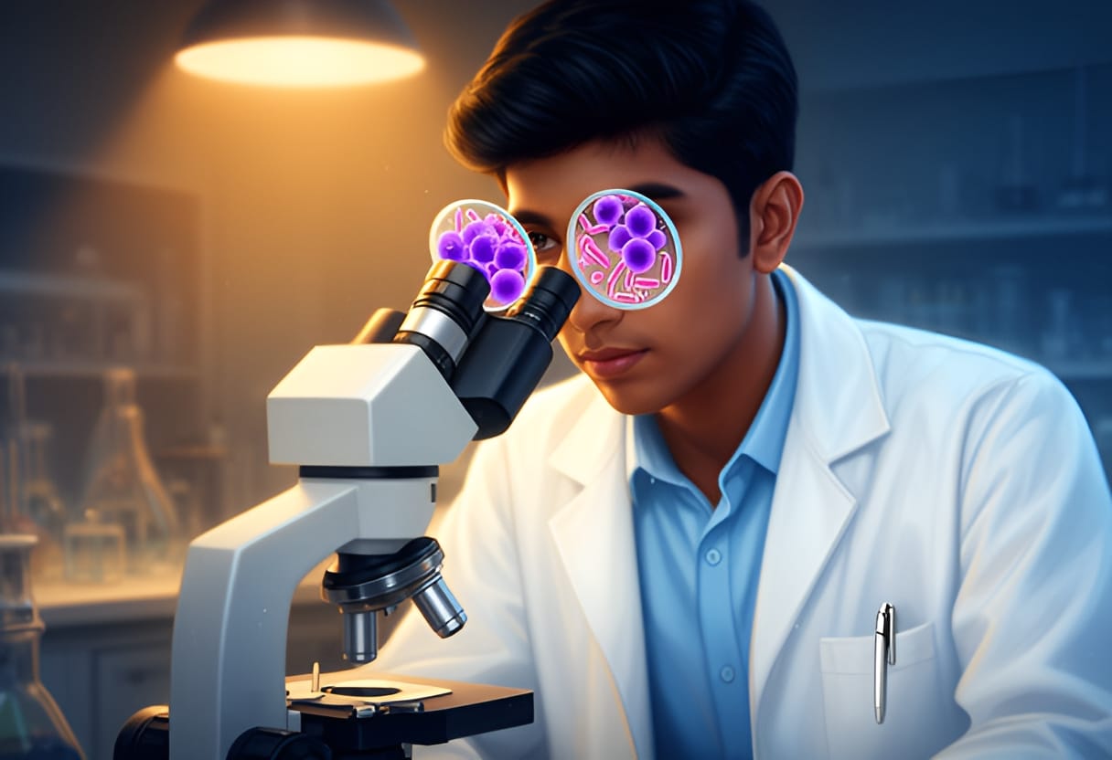
His breath hitched.
"Staph aureus is Gram-positive cocci," he murmured. "E. coli is Gram-negative rods."
Sneha leaned over. "You got it."
A grin spread across his face, unexpected and warm. He looked up to see Professor Meera observing from a few benches away, arms crossed, a quiet smile on her lips. She gave a nearly imperceptible nod.
Arjun lowered the microscope, the weight of the slide suddenly feeling less like a task and more like a promise. He hadn’t just followed steps. He had seen the difference—the wall that held the stain, the one that didn’t. And in that color, he saw not just bacteria, but a way to know them. To stop them.
Professor Meera set down a fresh slide labeled M. smegmatis beside Arjun’s successfully stained Gram slide. "Now we move to acid-fast staining," she said, her tone shifting to one of quiet intensity. "This technique targets organisms with waxy cell walls rich in mycolic acids—like Mycobacterium tuberculosis. Ordinary stains wash right out. But with heat and a lipid-soluble dye, we can trap the color inside, even after washing with acid-alcohol."
Arjun leaned forward as she demonstrated. She flooded the smear with carbol fuchsin, then held the slide over the Bunsen burner’s flame, not to boil, but to steam gently for five minutes. "Heat opens the waxy layer," she explained, "letting the dye penetrate. Then we decolorize with acid-alcohol—a mix of hydrochloric acid and ethanol. Most bacteria lose the stain. Acid-fast ones hold onto it. Finally, we counterstain with methylene blue to make the background visible."
She handed him the slide. "Your turn."
Arjun replicated the steps, careful with the flame, timing the steaming by counting slowly as Sneha had advised. When he applied the acid-alcohol, he watched anxiously. The red dye stayed put, clinging stubbornly to the cells. After rinsing, he added the methylene blue, waited, rinsed again, and blotted dry.
Through the microscope, the field glowed with bright red rods, some beaded, some branching slightly. The background was a pale blue.
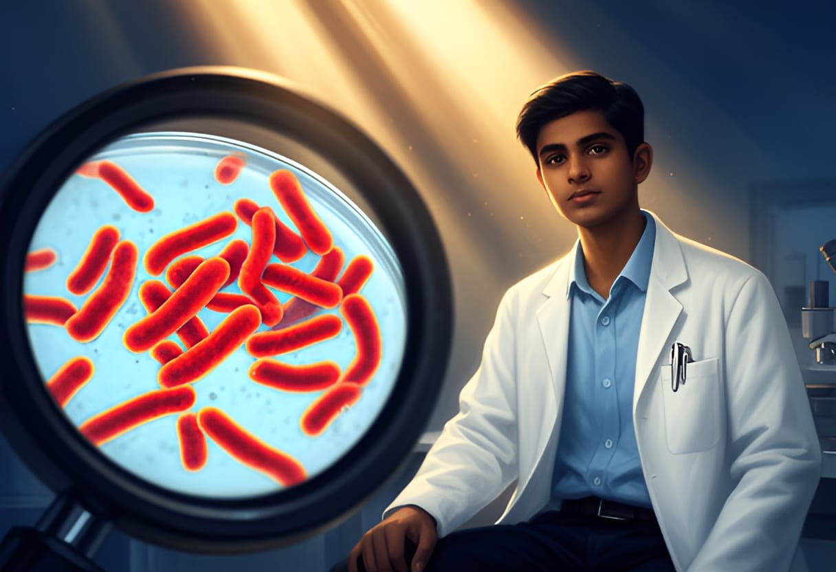
"Acid-fast," he whispered. "They’re red."
"Exactly," Professor Meera said. "And in pharmaceutical microbiology, we use this test not just for pathogens, but for environmental monitoring. Water systems, raw materials—especially those of natural origin—can harbor mycobacteria. They’re resistant to standard disinfection, so detecting them early prevents contamination of products like inhalants, irrigations, or even certain syrups where water is a component."
Arjun thought of the intravenous vials from Professor Meera’s first lecture, the ones that could cause sepsis. "So if we find acid-fast organisms in water used for injection, we know the system isn’t safe?"
"Precisely," she said. "It’s not about killing every microbe—that’s impossible. It’s about knowing what’s there, understanding its resistance, and acting before it compromises a sterile product. Staining gives us that knowledge. It turns the invisible into data."
Sneha pointed at the slide. "Look—some are clumped. Like they’re forming a biofilm."
Professor Meera nodded. "Observant. That’s another clue. Acid-fast organisms in clusters often suggest environmental persistence, not just transient contamination. It helps us trace the source."
Arjun held the slide up to the light, the red organisms stark against the blue. He thought of his initial question—How do we stop them?—and realized it wasn’t about stopping microbes entirely. It was about knowing them well enough to anticipate where they might hide, how they might survive, and where to intervene.
He looked up. Professor Meera was watching him, not with approval alone, but with the quiet satisfaction of someone seeing understanding take root.
Arjun smiled. The promise he’d felt after the Gram stain wasn’t just about seeing color. It was about seeing purpose. And now, with this new shade of red, the picture was clearer than ever.